• Home
  • Therapeutic Nuclear medicine

NUCLEAR MEDICINE

Nuclear medicine uses radionuclides both for   Diagnosis by Internal administration for therapeutic purposes.
nuclearmed2

Introduction

The department of Nuclear Medicine at the institute was started in November, 1957 headed by Dr.V.M.Sivaramakrishnan, M.A., Ph.D., D.R.I.P (Oak Ridge). Dr.Taylor, and Dr.V.K.Iya from Atomic Energy Establishment, Trombay visited this department to gather information for starting their Isotope Division. The Atomic Energy started supplying radioisotopes from 1961 onwards four years after the Nuclear Medicine department was started at Cancer Institute. Radioactive iodine and radioactive gold, imported from Harwell, UK were routinely used in the diagnosis and treatment of certain cancers. Dr.V.M.Sivaramakrishnan initiated major research programs involving the use of various radioisotopes with chelating agents in order to study the metabolic pathways in biological systems in both health and disease.

Scope

Nuclear medicine, related to oncology, nuclear oncology involves the uses of small amount of radioisotopes for study of organ function – functional imaging.

The availability of various radionuclides, have revolutionized the practice of medicine, during the last fifty years. Radionuclides have been successfully employed in clinical diagnosis and treatment. Though early progress was restricted by the limited choice of radio nuclides, by 1946 after the Second World War lot of isotopes was available for medical uses.

Radionuclides are used in four important ways in medicine (a) Diagnosis (b) Internal administration for therapeutic purposes (c) Brachytherapy and (d) Teletherapy sources for production of gamma ray beams.

The amount of radioactive material used weighs about 10-10 g; this means that studies can be carried out without any chance of the test method affecting the system being investigated.

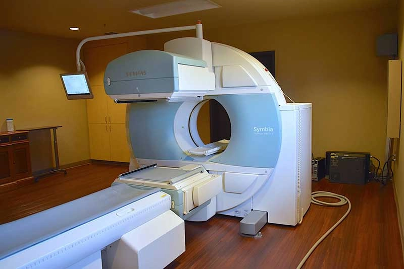
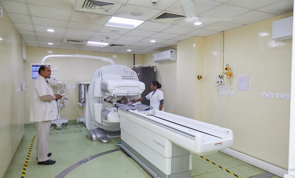
In nuclear medicine clinical information is derived from observing the distribution of a pharmaceutical administered to the patient. In-vivo imaging is the most common type of procedure in nuclear medicine, nearly all images being carried out with a gamma camera. It offers the potential, unique among imaging techniques, of demonstrating function rather than simple anatomy.

The presence of increased uptake of the radiopharmaceutical may demonstrate abnormal pathology, such as metastases several months before there has been sufficient bone erosion for the x-ray to appear abnormal in bone imaging. [read more]
The test which can be carried out with radionuclide can be broadly divided into three groups, namely metabolic and physiological studies, the measurement of body composition or body ‘spaces’ and organs and tumor localization and visualization.

In comparison to other diagnostic tests, the demonstration of physiological function makes a unique contribution to medicine. Thyroid function tests using I-131, imaging of the skeleton using Tc-99m labelled compounds, tumour imaging with F-18 (i.e.) fluorodeoxyglucose with PET scanner are some of the most commonly employed imaging procedures. Similarly the hepatobilary system, the gastrointestinal tract, the urinary track, vascular and lymphatic system can be investigated using Tc-99m labelled compound. [/read]

Emergency Cases

Please feel welcome to contact our friendly reception staff with any general or medical enquiry call us.

Opening Hours

Monday – Friday :  8.00 am – 7:00 pm
Saturday : 9.00 am – 8.00 pm
Sunday : 10.00 am – 9.00 pm

Registration Time

Monday – Sunday : 7.00 am – 11.00 am

Visiting Hours

Monday – Sunday : 4.00 PM – 6.00 PM

How It Began

To relate the beginning of nuclear medicine is to trace a convergence of many other scientific disciplines, with those of medicine, to illustrate the interdependence of basic science, medicine and technology and indeed to suggest that such electricism may well be a prophetic model of future advances in all fields of medicine.

Today the science and technology of nuclear medicine are used to solve problems involving every organ system of the body. The field can be thought of as “Topographic Physiological Chemistry” that is, in-situ chemistry. No field is better able to apply advances in molecular biology and genetics to the care of the sick and to biomedical research. In the perspective of the patients’ problem, the technological advances such as Positron Emission Tomography, Single Photon Emission Tomography, advanced image processing, information and large numbers of new physiological tracers, help in their solution.
Activities

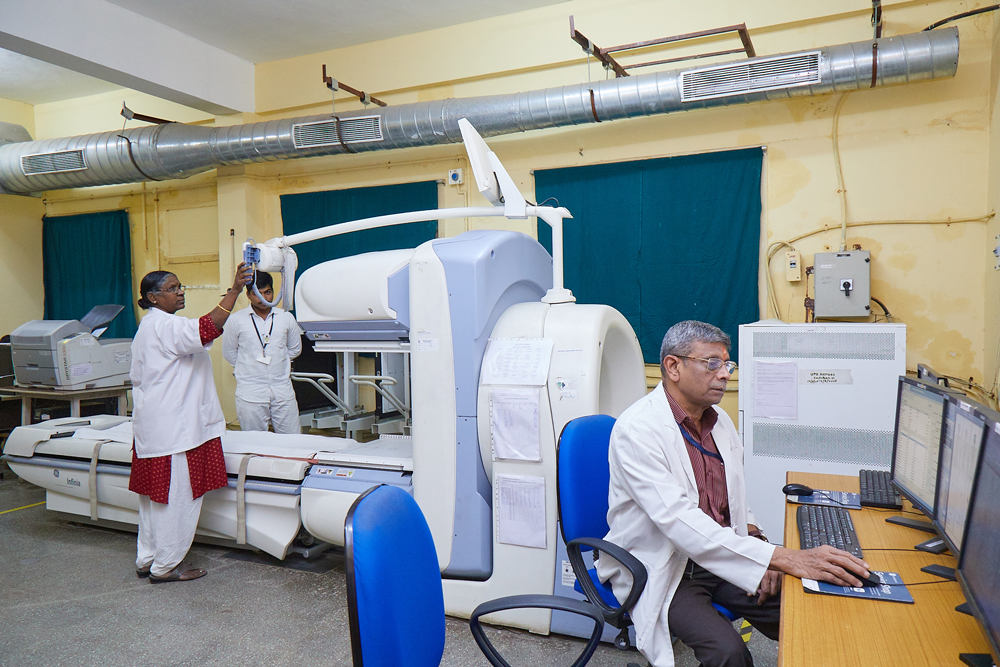
Doing bone scans, I-131 neck uptake and scans and whole body metastatic survey with I-131 for differential thyroid cancer, Ejection Fraction Calculation by MUGA technique, Renogram (Tc-99m DTPA) for evaluation of GFR , DMSA(V) scans for medullary carcinoma, I-131 MIBG scan for neuroectodermaltumours (Phaechromocytoma, Neuroblastoma etc.) and Ga-67 studies for lymphoma and Tc-99m MIBI for Parathyroid Scintigraphy and Scintimammography, SPECT for haemangioma (Tc-99m RBC studies) Ventilation Scan (Tc-99m DTPA) and Perfusion Scan (Tc-99m MAA) for prediction of postoperative FEV1 calculation in lung resection. Therapeutic nuclear oncology includes high dose therapy for Ca.Thyroid with I-131, Radio phosphorous and Radio Samarium for palliation of bone metastases, I-131 MIBG therapy for neuroendocrine tumours.

The amount of radiopharmaceutical used is carefully selected to provide the least amount of radiation exposure to the patient but ensure an accurate test

Nuclear Medicine studies involve the use of small amount of radioactive isotopes administered into the body.

It is mainly used to detect the spread of cancer to the bones especially in cancer of breast, prostate, thyroid & lung.

The functions of organs like kidney, liver and brain are also being studied by the isotopes.

We also use similar isotopes for studying the function of thyroid gland.

Radioactive iodine is used to detect the spread of cancer into other organs.

We also use such isotopes for the treatment of the similar spread to other organs.

Cost effective.

Nuclear medicine is practiced only by

The various types of NM images can be classified as

Radiation safety

CMEs are conducted by the department. Invited speakers from reputed institutions in the country and from abroad participate.

Academic programs conducted

Observership Training given for post graduates of M.Sc [Medical physics] of Govt.Arignar Anna Cancer Hospital, Karapettai, Kanchipuram in our nuclear medicine department for a period of 1 month in 2 batches from since 2013 as a part of their Internship programme.

Ongoing Project

The Study of Global Methylation and Braf mutations in Papillary Thyroid cancers – Role in modulating Radio ablation by I-131

Future Projects

Randomized controlled trial of I-131 Tositumomab (Bexxar) radio-immunotherapy versus rituximab immunotherapy with relapsed or refractory low grade, follicular or transformed B-cell Non-Hodgkin’s Lymphoma.

Staff

NAMEEDUCATIONAL QUALIFICATIONDESIGNATION
Dr. R. Krishna kumarM.D., DMRT, DRM, Ph.DProfessor and Head
Mrs. A. Balkis BegumM.Sc[ Medical Physics].,Medical physicist cum RSO(III)
Mr. G.K. RangarajanM.Sc., ANMT, RSOScientific Assistant cum Technologist
Mrs. N.AnandiDMLT, B.Sc [NMT]Technician
Mr.Kuncha RameshDMLTTechnician

Students

NAMEEDUCATIONAL QUALIFICATIONDESIGNATION
Mr.N.Parvathi NathanB.sc N.M.T (3rd Year)Student
Miss.S.LavanyaB.sc N.M.T (3rd Year)Student
Mr.V.SandilyanB.sc N.M.T (2nd Year)Student
Miss.D.DivyaB.sc N.M.T (2nd Year)Student
Miss.SyedAzheemunnisaB.sc N.M.T (1st Year)Student
Miss.U.JayanthiB.sc N.M.T (1st Year)Student
Mr.R.SridharB.sc N.M.T (1st Year)Student
Mr.H.GokulDassB.sc N.M.T (1st Year)Student


Publications

  1. Prediction of postoperative pulmonary reserve in lung resection patients published in polish journal of Nuclear Medicine , 2011

2. Krishnakumar,R. Priyadarshini,R..Manivannan, K.Shanmugasundaram,B. and.Anuradha (2003) Quantification of Right to Left Shunt by Tc-99m Macro aggregated Albumin-A case report. Indian Journal of NuclearMedicine, Vol. 18 No.3, pp 73-75

3. R.Krishnakumar, R.Priyadarshini, K.Elumalai, G.K.Rangarajan, S.Ayyappan, SarojiniPrahalad, VandhanaMahajan, A.V.Lakshmanan,Ossifying Metastases from Osteosarcoma of Left Humerus Demonstrated by Bone Scintigraphy.Indian Journal of Nuclear Medicine Vol 21, No.3, pp 69-71 Sep’2006

4. Co-author for a case report ESOPHAGEAL METASTASES FROM CARCINOMA OF THYROID published in IJNHS ,2011

5. R.Krishnakumar, N.Nair, K.Makhdoni et al.”Tetrfosmin Imaging in Lymphoma”,ANNALS OF ONCOLOGY, Vol.10, 1999, Abstract for Publication in 7th International meeting of Lymphoma, Switzerland

6. Krishnakumar, R. (2002) Prediction of vascular insufficiency in anticipated large vessel Sacrifice (Abstract) World Journal of NuclearMedicine,Vol.1, (Supplement I), p S 82

7. Krishnakumar, R, Priyadarshini.R, Balkis Begum (2003) Comparison of Gallium-67 and Tc-99mTetrofosmin uptake in lymphoma (Abstract) , Bangladesh journal of Nuclear Medicine,Vol-6, No.1, Pg 58, 2003.

8. Krishnakumar, R. Priyadarshini.R, Balkis Begum (2003) Assessment of response of limb sarcoma to neoadjuant chemotherapy with Tc99m Sesta MIBI, (Abstract), Bangladesh journal of NuclearMedicine, Vol-6, No.1, Pg 4, 2003 .

9. Krishnakumar,R. Priyadarshini, R and Bulkis Begum (2004) “Comparison of Ga-67 and Tc-99m Tetrofosmin uptake in lymphoma”(Abstract), World Journal of NuclearMedicine, Vol.3 (Supplement I), p 34

10. Krishnakumar, R. Priyadarshini,R. Shanmugasundaram,B. Bulkis Begum and. Anuradha (2004) Assessment of response of limb sarcoma to neoadjuant chemotherapy with Tc-99m Sesta MIBI” Abstract), World Journal of NuclearMedicine,Vol.3( Supplement I), p 93.

11. Krishnakumar.R, A.V.Lakshmanan, Priyadarshini.R,Elumalai and Rangarajan, “Prediction of Post Operative Pulmonary reserve in Patients who undergo Lung Resection” at the national Annual AMPI conference, Journal of Medical Physics, 30, 4, (2005) Pg.287.

12. Veeraiah S, Elangovan V, Tripathy JP, …, Rajaraman S, … et al. Quit attempts among tobacco users identified in the Tamil Nadu Tobacco Survey of 2015/2016: a 3-year follow up mixed methods study. BMJ Open 2020; Sep 3 10(9): e034607 PMID: 32883722

13. Role of Scintimammography in the evaluation of response to neo-adjuvant chemo-radiotherapy in cancer of breast, IJNM, Sep 2007, Vol.22, No.1

14. “Characterization of a new MOSFET detector configuration for in vivo skin dosimetry”,PaoloScalchi, Paolo Francescon, and Priyadarshini Rajaguru, Department of Medical Physics, San Bortolo Hospital, Vicenza, Italy,Med. Phys. 32 (6), Pg 1571-1578, June 2005, An official journal of American Association of Medical Physicists and Canadian Association of Medical Physicists.

15. Priyadarshini Rajaguru, co-author to a paper presented on “Application of Radiobiological model in Intensity modulated radiation therapy (IMRT)”,PaoloFrancescon, Stefania Cora, Michele Avanzo, Priyadarshini Rajaguru, RosabiancaGuglielmi, Gino Panizzoni ,at Legnaro-Padova, during the period 20-22 novembre 2003.

16. Published in Proceedings of II Reunion of National Society of Italian Research in Radiation, I meeting of National Federation of Italian research in Radiation, under the topic, ‘Radiation in Medicine and Biology: status of research in clinical applications’, ‘RadiozioniRicerca e Applicazioni’ Suppl. Vol VII no.1 Bollettinodella SIRR Aprile 2004 Pg 57-59.

17. Indian Journal Of Nuclear Medicine June 2006 VOL.21 No.2, IJNM, 21(2): 32 – 35,2006, Comparison of Gallium -67 and Tc-99m Tetrofosmin Uptake in Lymphoma, Dr.R.Krishnakumar, Dr.R.Priyadharshini, Bulkis Begum.

18. IJNM, 21(2) :45 – 46,2006, Ureteric Activity Mimicking Bone Uptake in Tc-99m MDP Dr.R.Krishnakumar, Dr.R.Priyadharshini,K.Elumalai,G.K.Rangarajan and Dr.A.VLakshmanan

19. Indian Journal Of Nuclear Medicine September 2006 Volume 21 No:3,Assessment of Response of Limb sarcoma’s to Neoadjuvant Chemotherapy with Tc99m Sesta MIBI , R.Krishnakumar, R.Priyadarshini, B.Shanmugasundaram, Bulkis Begum,Anuradha, A.V.Lakshmanan, S.Ayyappan, U.Majhi

20. Case Report IJNM , 21(3) :69 – 71 ,2006 Sep 2006 Volume 21 No :3, Ossifying Metastases from Osteosarcoma of Left Humerus Demonstrated by Bone Scintigraphy, R.Krishnakumar, R.Priyadarshini, K.Elumalai, G.K.Rangarajan, S.Ayyappan, SarojiniPrahalad, VandhanaMahajan, A.V.Lakshmanan

21. Case Report (WJNM)World Journal of Nuclear Medicine , 201 – 204,July.2006, Vol-5, No:3,Paget’s disease and Bone Scan, R.Krishnakumar, R.Priyadarshini, K.Elumalai, G.K.Rangarajan, S.Ayyappan,

22. Exploring the role of technitium-99m dimercaptosuccinyl acid (V) scan in medullary carcinoma thyroid patients with postoperative persistent hypercalcitoninemia in the era of positron emission tomography-computerized tomography, IJNM,2014.Aravinth Krishnamurthy,R.Krishnakumar, R.Praveen, R.Vijayalakshmi, A.Balkis Begum, G.K.Rangarajan.

    Make Appointment

    Call: 044-22209150/22350131 Ext 105/152/164/169/211 between 7AM and 5PM

    Quick Contacts

    Please feel free to contact our friendly staff with any medical enquiry.

    Health Essentials

    News & Articles

    Flyer YHM-2024

    Youth Health Mela 2024

    Join the Movement: Empowering Youth for a Healthier Tomorrow! The mission is to engage, educate, and inspire young minds to embrace a healthier lifestyle through increased physical activity. Come &…
    Read more