

Radio Diagnosis and Imaging
The Department of Radiology focuses on advanced imaging and image-guided procedures for accurate cancer diagnosis, staging, and treatment planning.
radiology@cancerinstitutewia.orgAbout the department
The Department of Radiology at Cancer Institute (W.I.A) provides comprehensive imaging services that play a vital role in the diagnosis, staging, treatment planning, and follow-up of cancer patients.
Equipped with advanced imaging technologies and supported by a team of experienced radiologists and technologists, the department offers a wide range of diagnostic and image-guided interventional procedures. These services include X-Ray, Ultrasound, Mammography, Computed Tomography (CT), Magnetic Resonance Imaging (MRI), and image-guided biopsies and therapeutic interventions.
The department works in close coordination with oncology, surgery, radiation therapy, and other clinical specialties to ensure accurate diagnosis and optimal patient care. Emphasis is placed on patient safety, adherence to protocols, and maintaining high standards of quality in imaging and reporting.
Every effort is made to provide a comfortable and supportive environment, with clear communication and guidance at each step of the imaging process to aid diagnosis.
Message from the Head of Department
“The Department of Radiology is committed to delivering accurate, timely, and safe imaging services that form the foundation of effective cancer care. Working in close collaboration with medical, surgical, and radiation oncology teams, the department plays a vital role in diagnosis, staging, treatment planning, and follow-up. With a strong emphasis on precision, safety, and patient comfort, the team integrates advanced imaging technology with evidence-based practice to support the Cancer Institute’s mission of compassionate, patient-centered, and multidisciplinary oncology care . ”
Dr. Karthigaiselvi M
Senior Consultant Radiologist & Incharge - Department of Radio Diagnosis & Imaging
Diagnostic techniques and procedures
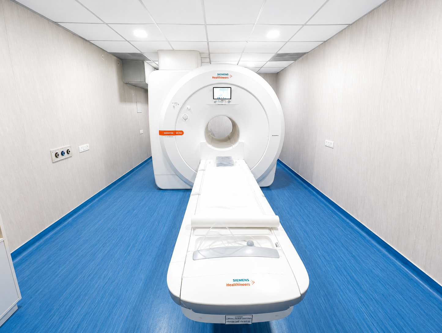
MRI (Magnetic Resonance Imaging)
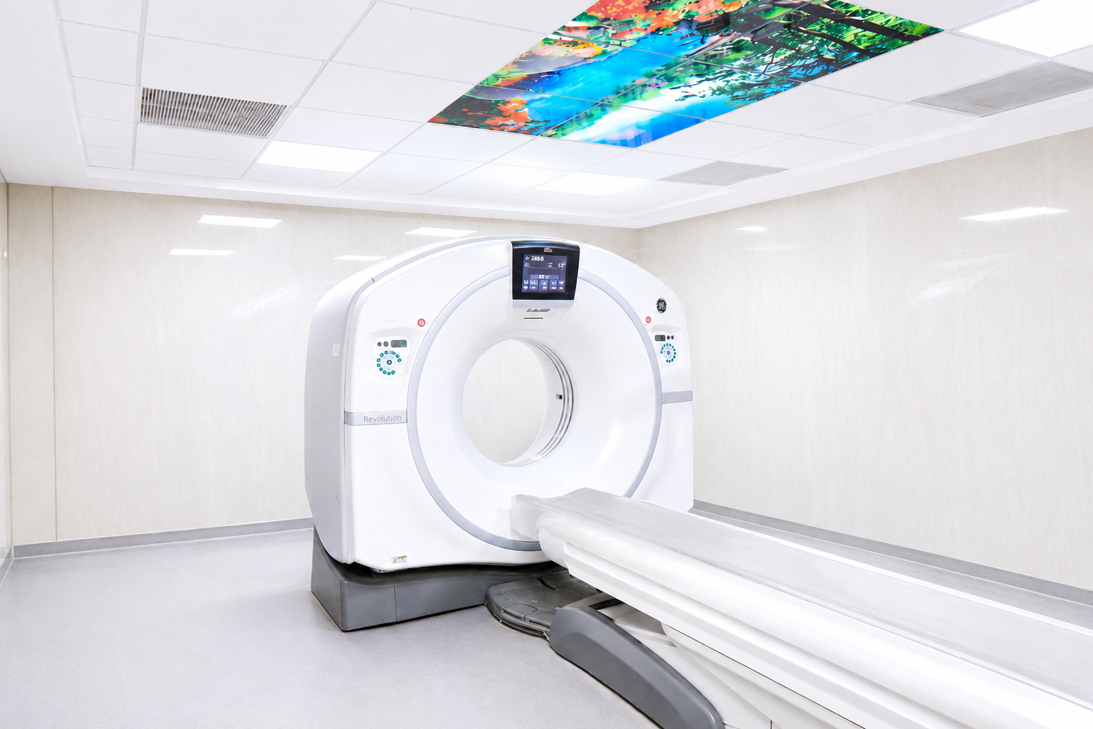
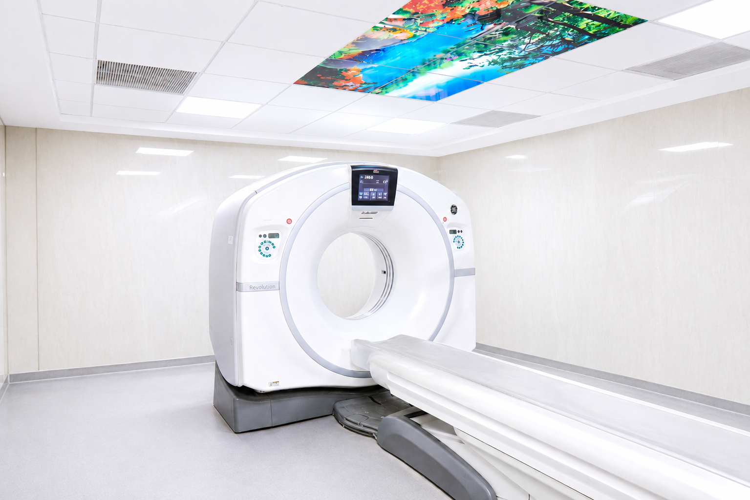
CT Scan (Contrast and Non-Contrast)
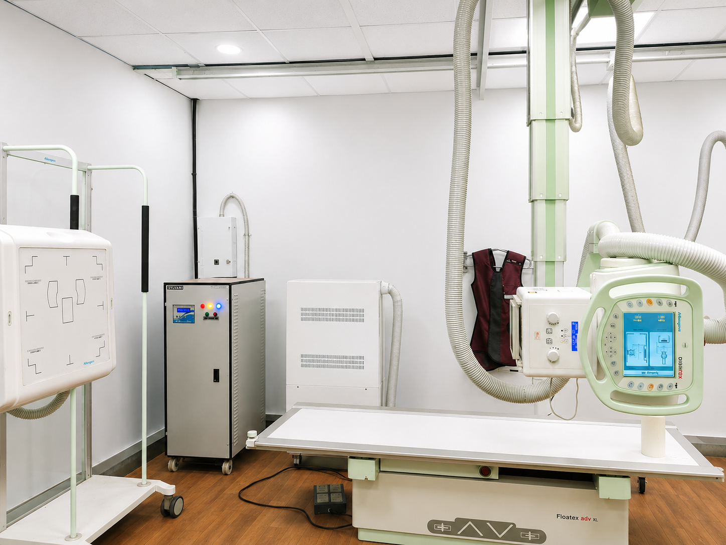
X-ray
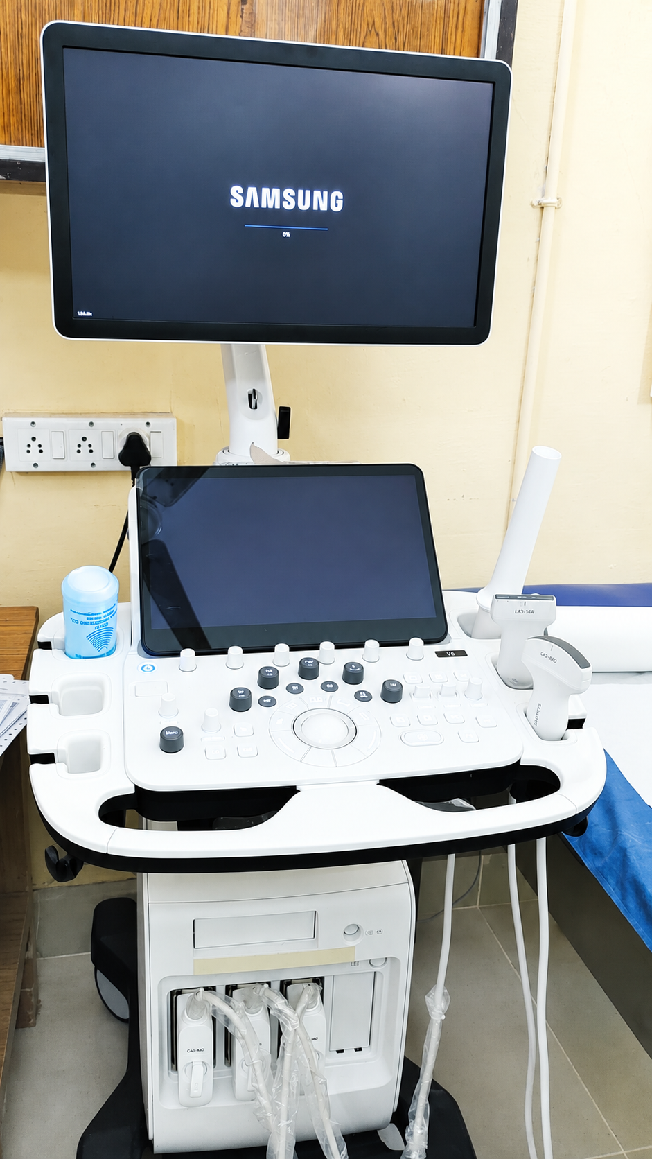
Ultrasonography (USG)
Transvaginal Ultrasound (TVS)
Transrectal Ultrasound (TRUS)
Thyroid Ultrasound
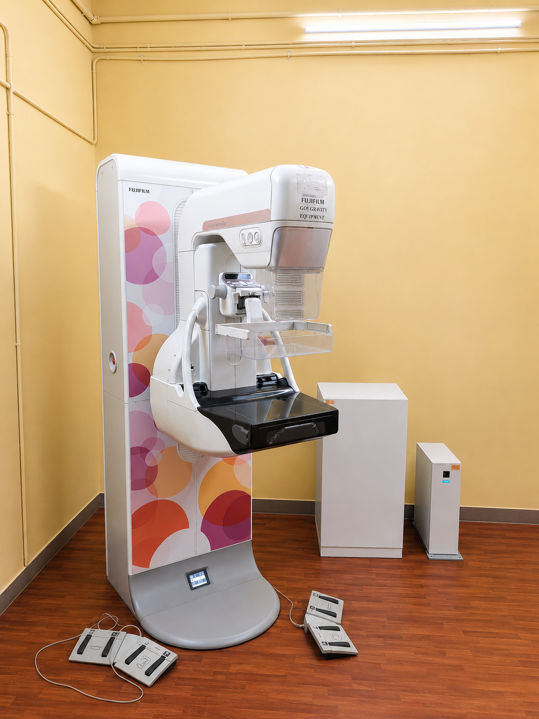
Mammography
Digital Breast Tomosynthesis (Tomo)
Contrast-Enhanced Mammography (CE Mammography)
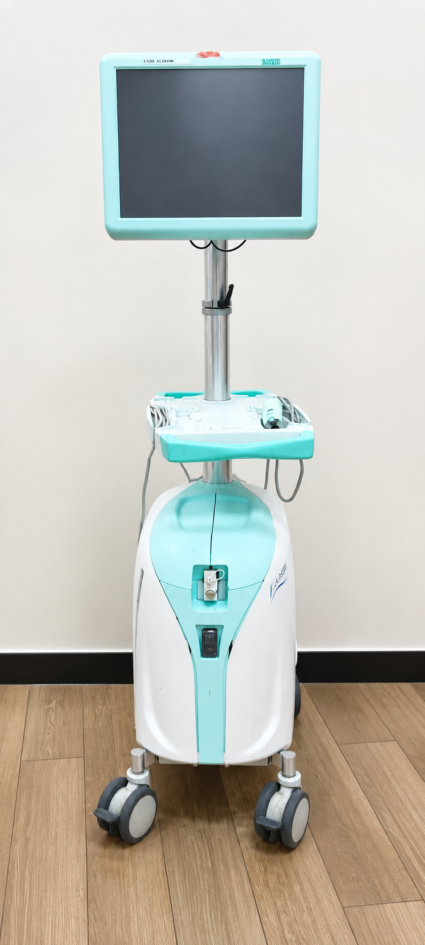
Stereotactic Biopsy
Ultrasound-Guided Procedures
Vacuum-Assisted Biopsy (VAB)
CT-Guided Procedures
Fiducial Marker Placement
J-Wire Localization
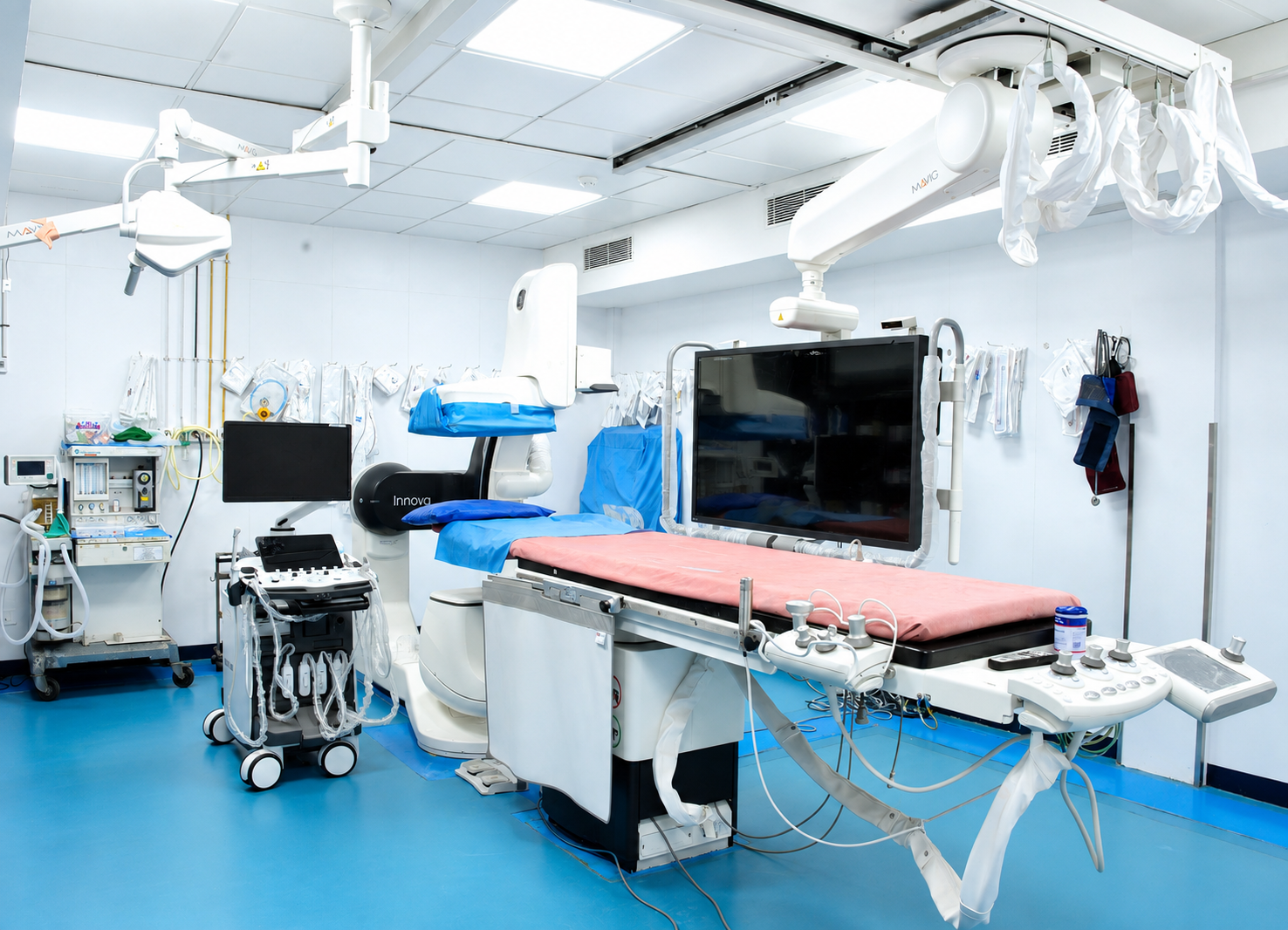
Cathlab Diagnostic Procedures
Cathlab Therapeutic Procedures
GENERAL INFORMATION
Preparing for Your Visit
Everything you need to know before, during, and after your radiology appointment at Cancer Institute (W.I.A), Adyar.
What You Should Bring
- Previous scans and reports
- Doctor's referral
- Previous imaging (CT/MRI/X-ray films or CDs)
- Reports and discharge summaries
- Blood test reports (if advised, especially renal function)
Information You Should Share with Us
- Pregnancy or possibility of pregnancy
- Allergies (especially contrast)
- Kidney disease or diabetes (especially on insulin/metformin)
- Implants (pacemaker, stents, clips, prosthesis)
- Claustrophobia
- Current medications (blood thinners)
During Your Visit
- Arrive 30–60 minutes early
- Fasting may be required (depends on test)
- Wear loose, metal-free clothing
- Follow staff instructions during scan
- Remain still when required
- You will be guided by trained staff
- Some scans require contrast injection
After Your Test
- Most tests: no restrictions
- Some procedures require short observation
- Drink fluids if contrast used
Turnaround Time (TAT)
- Routine reports: 24–48 hours
- Urgent cases: same day (if indicated)
Consent
(Standard hospital practice)
- You may be asked to sign a consent form
- Procedure will be explained beforehand
When to Seek Help After a Procedure
Contact hospital immediately if:
- !Severe pain
- !Bleeding
- !Breathlessness
- !Rash or swelling after contrast
PATIENT SAFETY & CLINICAL PROTOCOLS
Radiology Safety Guidelines & Patient Care
At Cancer Institute (WIA), patient safety, comfort, and clinical precision are our highest priorities. Explore our comprehensive imaging safety protocols, special situation advisories, and aftercare instructions below.
Special Situations
Important conditions to disclose to your care team prior to scanning
Pregnancy
- Please inform the radiology team if you are pregnant or suspect pregnancy
- Certain scans (especially CT) may be postponed or modified
- Alternative imaging methods may be used when appropriate
Patients with Diabetes
- Inform if you are on insulin or oral medications (especially metformin)
- Instructions regarding fasting and medication adjustment will be provided
Patients with Implants or Devices
- Inform about pacemakers, stents, clips, prostheses, or metal implants
- This is especially important for magnetic resonance (MRI) scans
Children and Elderly Patients
- Additional care and assistance will be provided
- A caregiver may be allowed to accompany the patient when necessary
Understanding Contrast Reactions
Contrast dye is safe and well tolerated by the vast majority of patients. Understanding expected sensations helps ensure a calm and prepared visit.
Common Mild Reactions
These are usually temporary and resolve on their own.
Rare Reactions (Seek Immediate Help)
Our team is trained to manage such reactions promptly.
Aftercare & Warning Signs
Post-procedure guidance and monitoring for our patients
After Imaging with Contrast
- Drink plenty of fluids to help eliminate contrast
- Resume normal diet unless advised otherwise
After Image-Guided Procedures
- Mild pain or discomfort at the site may occur
- Avoid strenuous activity for 24 hours
Contact the Hospital Immediately If You Experience
- Severe or increasing pain
- Bleeding or swelling at procedure site
- Fever
- Breathing difficulty or rash
Patient Comfort & Support
We understand that medical tests can sometimes be stressful.
Treatment options
- Image-Guided Biopsies
- Interventional Radiology Procedures
- Tumor Embolization
- Microwave ablation
- Drainage Procedures
- Stent placements
- Fiducial Marker Placement
- Vascular Access Procedures
Preventive measures
Mammography Screening
Women over 40 years should undergo annual or biennial mammograms for early detection of breast cancer.
Ultrasound (USG) Screening
Recommended for abdominal, pelvic, thyroid, and breast evaluation in selected high-risk individuals.
High-Risk Breast Screening
Additional imaging such as ultrasound or MRI may be advised for women with dense breasts or strong family history.
Surveillance for Cancer Recurrence
Regular follow-up imaging (X-Ray, CT, MRI, USG, or Mammography) as advised by your oncologist Imaging to monitor treatment response Early detection of recurrence or metastasis through scheduled scans
When to visit
- If a doctor advises imaging for suspected cancer
- If you notice a new lump in the breast or any part of the body
- If you have persistent headaches, seizures, or neurological symptoms
- If you experience unexplained weight loss
- If you have persistent cough or chest pain
- If there is abnormal bleeding or discharge
- If follow-up scans are required after cancer treatment
- If routine cancer screening is due
Legacy of the department
- The Department of Radiology at the Cancer Institute (W.I.A.), Adyar, has been a vital component of the Institute’s cancer care services since its formative years. As one of India’s pioneering comprehensive cancer centres, the Institute recognized early the importance of accurate imaging in diagnosis and treatment planning. Beginning with conventional X-ray services, the department steadily expanded in scope and capability, keeping pace with technological advancements in cancer imaging.
- The department was part of the Institute’s early adoption of mammography in the 1960s, contributing to improved early detection of breast cancer in India. Over time, Radiology evolved into a comprehensive imaging and interventional specialty, incorporating ultrasound, CT (contrast and non-contrast), MRI, advanced mammography techniques, and image-guided diagnostic and therapeutic procedures.
Key achievements
11 Milestones- 01National Board of Examinations (NATBOARD) Recognition for DNB Radiology Training Programme – Ongoing – 2023
- 02National Board of Examinations in Medical Sciences (NBEMS)
- 03ICRI Fellowship in Onco-Imaging: Initiated 2024 – International Cancer Research Initiative (ICRI)
- 04ICRI Fellowship in Breast Imaging: Initiated 2024 – International Cancer Research Initiative (ICRI)
- 051st Prize: Oral Paper Presentation – 2024 – National IRIA Conference, Vijayawada
- 06Prize Winner: RITE National Conference (Quiz & Oral Paper Categories) – 2025
Our Doctors

Dr. Karthigaiselvi M
Senior Consultant Radiologist & Incharge - Department of Radio Diagnosis & Imaging
View profile
Dr. R. Thanraj
Consultant Radiologist and Sonologist
View profile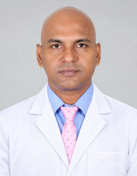
Dr. Pushparajan S.
Intervention Radiologist
View profileDr. Anusuya P
Consultant Radiologist
View profileDr. Vignesh M.
Consultant Radiologist
View profileDr. Revathy P.
Consultant Radiologist
View profile
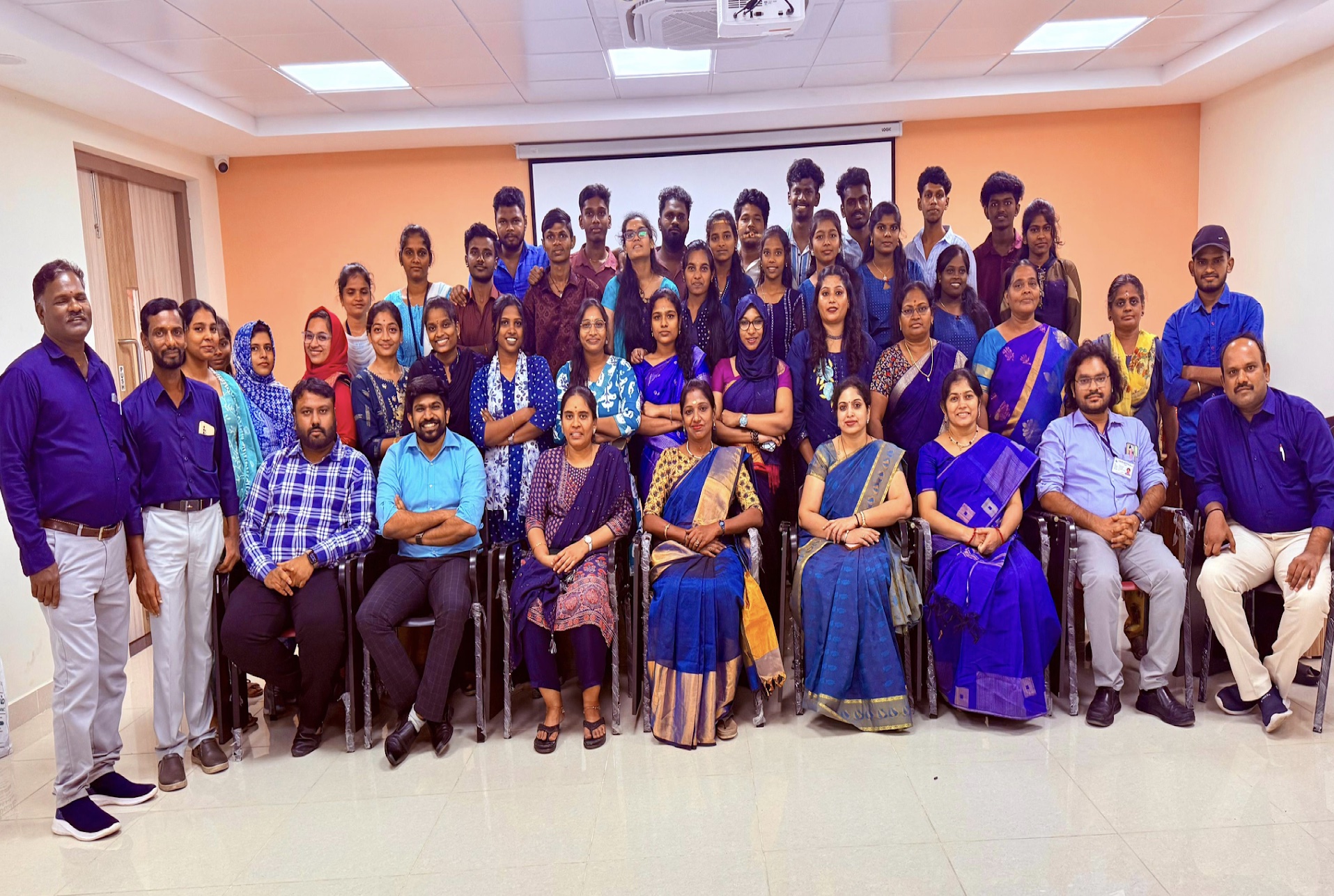
Our Team
Patient guidance
Frequently asked questions
Will the scan be painful?
Most imaging tests are painless. Some procedures or contrast dye injections may cause mild, temporary discomfort.
Can I eat before my scan?
Contrast MRI, CT scans, and interventional procedures require 4–6 hours fasting. Abdominal ultrasound requires 6–8 hours fasting. Plain X-rays and soft tissue ultrasound need no fasting.
How long will the test take?
Most diagnostic imaging scans take 10 to 45 minutes. Interventional biopsies and Cathlab procedures take 20 to 60 minutes plus 2–4 hours post-procedure observation.
When will I get my report?
Routine imaging reports are available within 24–48 hours. Urgent cases are reported the same day when indicated.
Can I go home immediately after the test?
Yes, most diagnostic patients resume normal activities immediately. Interventional biopsy and Cathlab patients undergo 2–4 hours of observation before discharge.
What should I bring for my visit?
Bring your doctor's referral letter, previous scans/films/CDs, discharge summaries, and blood test reports (especially Serum Creatinine for contrast scans).
Is radiation safe during scans?
CT and X-ray examinations follow the ALARA principle—using the lowest radiation dose required for accurate diagnosis. MRI and Ultrasound use zero radiation.
How are contrast reactions managed?
Contrast dye is generally safe. A warm sensation or metallic taste is normal. Allergic reactions are rare, and our radiology team is fully trained with emergency protocols.
How is patient privacy protected?
All patient information and imaging results are kept strictly confidential and shared only with the treating multidisciplinary oncology team.