#01
Earlier detection
identifies cancer and disease at the molecular level, often years before structural changes appear on CT or MRI
Clinical Advantage
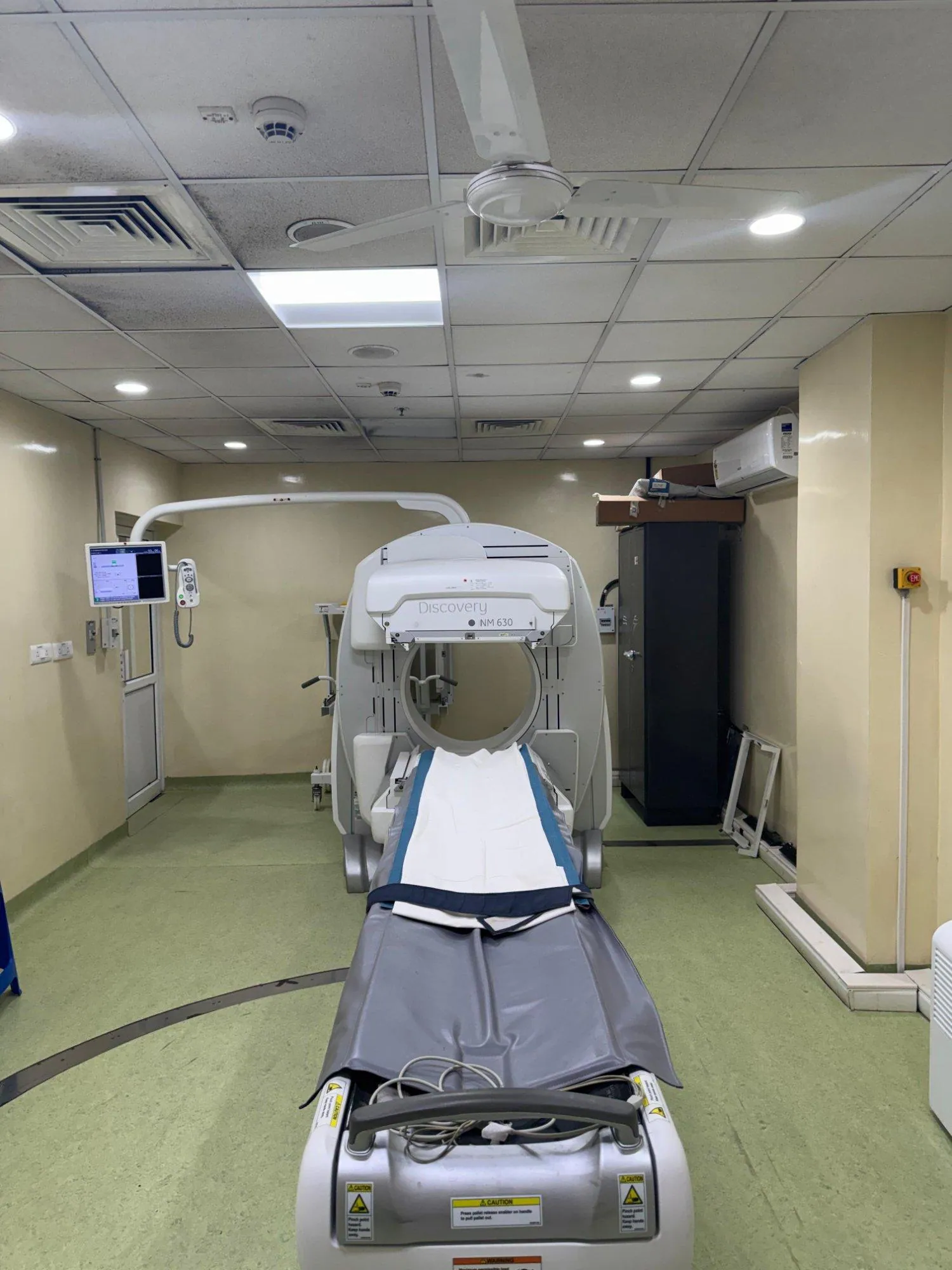
Nuclear medicine focuses on diagnosing and treating cancer and other diseases using radioactive substances.
nm@cancerinstitutewia.orgSee disease from the inside — nuclear medicine & molecular imaging Unlike X-rays or MRI which show anatomy, nuclear medicine scans reveal how your organs and tissues are functioning at the molecular level — detecting disease earlier and with greater precision than any other imaging method.
We carry forward a legacy that began in 1957 when our founding pioneer, Dr. V.M. Sivaramakrishnan, established one of India’s first nuclear medicine centers. Today, we honor that vision by combining our six decades of experience with cutting-edge molecular imaging and therapeutic technologies. Our department stands at the unique intersection of physics, chemistry, and medicine—using the power of radiopharmaceuticals to see what others cannot see and treat what others cannot reach. Every nuclear medicine scan, every radionuclide therapy, and every diagnostic procedure we perform is driven by one commitment: to provide our patients with the most accurate diagnosis and effective treatment possible. We have a dedicated team of physicians, physicists, and technologists who work tirelessly to ensure that each patient receives personalized, evidence-based care with compassion and dignity.
Dr. Ashok Kumar R
Consultant & Incharge - Department of Nuclear Medicine
MOLECULAR INSIGHT
Established in November 1957, our department is one of India's pioneering nuclear medicine centers. We were among the first to use radioactive iodine and radioactive gold for cancer diagnosis and treatment - even before the Atomic Energy Establishment began regular isotope supply in 1961. Our founder, Dr. V.M. Sivaramakrishnan, initiated groundbreaking research on radioisotopes and metabolic pathways. Today, we continue this legacy with state-of-the-art gamma cameras and advanced therapeutic nuclear medicine capabilities.
WHAT MAKES IT DIFFERENT
Conventional scans — CT, MRI, ultrasound — create detailed pictures of the body's structure. Nuclear medicine imaging goes further: a small amount of a radioactive tracer is introduced into the body and travels to the specific organ, tissue, or cell type being studied. A special camera detects the radiation emitted, creating images that show biological activity in real time.
This means cancer can be detected before a tumour is visible on CT, heart disease can be assessed without invasive procedures, and the spread of disease throughout the entire body can be mapped in a single scan.
WHY IT MATTERS
identifies cancer and disease at the molecular level, often years before structural changes appear on CT or MRI
maps disease throughout the entire body in a single session, enabling complete cancer staging without multiple procedures
confirms whether a tumour is metabolically active, identifies the best biopsy site, and monitors response to therapy
delivers targeted diagnostic information with radiation doses similar to a few months of natural background exposure
assesses organ function (heart, kidney, thyroid, brain) that cannot be obtained from structural imaging alone
OUR IMAGING MODALITIES
We offer the full range of nuclear medicine and hybrid imaging technologies, each suited to specific clinical questions.
Positron Emission Tomography / Computed Tomography
Combines functional PET imaging with detailed CT anatomy in a single session. The gold standard for cancer staging, restaging, and treatment response assessment.
Uses: Oncology, neurology, cardiology
Positron Emission Tomography / Magnetic Resonance Imaging
Next-generation hybrid combining PET with the superior soft-tissue contrast of MRI. Ideal for brain, liver, pelvis, and paediatric imaging with lower radiation dose.
Uses: Brain tumours, prostate, paediatric
Single Photon Emission Computed Tomography / CT
3D functional imaging fused with CT for precise localisation. Widely used for bone scans, cardiac perfusion, and endocrine tumour imaging. Excellent availability and cost-effectiveness.
Uses: Bone scan, cardiac, thyroid, renal function
Prostate-Specific Membrane Antigen PET/CT
Highly sensitive imaging of prostate cancer using PSMA-targeted tracers (Ga-68 or F-18). Detects recurrence at PSA levels as low as 0.2 ng/mL — far earlier than conventional imaging.
Uses: Prostate cancer staging and recurrence
Ga-68 DOTATATE / DOTATOC PET/CT
The most sensitive scan for detecting neuroendocrine tumours (NETs) throughout the body. Also used to select patients for PRRT therapy and to monitor treatment response.
Uses: Neuroendocrine tumours, PRRT selection
F-18 Fluorodeoxyglucose PET/CT
The most widely used PET scan. FDG is a glucose analogue taken up by metabolically active cancer cells, infection, and inflammation — enabling whole-body disease mapping in one scan.
Uses: Lymphoma, lung cancer, infection, inflammation
Myocardial Perfusion Imaging (MPI) · Viability · MUGA
Evaluates blood flow to heart muscle, identifies hibernating myocardium, and assesses heart function. Essential before revascularisation and for monitoring cardiotoxicity of cancer treatments.
Uses: Coronary artery disease, heart failure
Amyloid · Tau · FDG · Dopamine (DaTscan)
Detects Alzheimer's plaques, tau tangles, and dopamine transporter deficits years before symptoms worsen. Distinguishes Parkinson's disease from other movement disorders with high accuracy.
Uses: Dementia, Parkinson's disease, epilepsy
KEY RADIOTRACERS
Each tracer is designed to target a specific biological process, receptor, or cancer type. The choice of tracer determines what the scan can see.
| Tracer | Scan type | What it targets | Main use |
|---|---|---|---|
| F-18 FDG | PET/CT | Glucose metabolism | Most cancers, infection, inflammation |
| Ga-68 PSMA | PET/CT | PSMA receptor | Prostate cancer staging and recurrence |
| Ga-68 DOTATATE | PET/CT | Somatostatin receptor | Neuroendocrine tumours |
| F-18 NaF | PET/CT | Bone turnover | Bone metastases, skeletal survey |
| Tc-99m MDP | SPECT/CT | Bone metabolism | Bone scan — metastases, fractures, Paget's disease |
| Tc-99m Sestamibi | SPECT/CT | Myocardial perfusion | Cardiac stress test, parathyroid imaging |
| Tc-99m DTPA | SPECT/CT | Renal function | To evaluate renal GFR |
| Tc-99m HIDA | SPECT/CT | Liver function | Liver function, biliary leakage, biliary system evaluation |
| Tc-99m Sulphur colloid | SPECT/CT | Lymphatic system | Sentinel lymph node evaluation |
| F-18 Florbetapir | PET/MRI | Amyloid plaques | Alzheimer's disease diagnosis |
| I-123 MIBG | SPECT/CT | Adrenergic tissue | Phaeochromocytoma, paraganglioma |
| F-18 FDOPA | PET/CT | Dopamine synthesis | Brain tumours, NETs, movement disorders |
WHAT TO EXPECT
Most nuclear medicine scans follow a similar pathway, with preparation instructions tailored to your specific tracer and clinical question.
Your doctor refers you to our department. Our team reviews the request, selects the appropriate tracer, and contacts you to confirm preparation instructions (fasting, medications to hold, hydration).
On arrival, the radiotracer is administered — usually by a simple intravenous injection, occasionally by inhalation or capsule. The dose is tiny and carefully calculated for your body weight.
You rest quietly for 30-90 minutes while the tracer distributes to the target tissue. For FDG scans, you will rest in a quiet, warm room to minimise background muscle activity. Uptake time varies for SPECT/CT images based on type of tracer used.
You lie comfortably on the scanner bed while images are acquired. Most whole-body PET/CT scans take 20-30 minutes. SPECT/CT imaging time varies based on type of scan performed.
A specialist nuclear medicine physician analyses your images and issues a detailed report to your referring doctor. Results are typically available within 48 hours. Our team is happy to discuss findings with patients directly.
PET/CT VS SPECT/CT
Both technologies have important roles. Your nuclear medicine physician selects the best modality for your clinical question based on the target tissue, required sensitivity, and available tracers.
DIAGNOSTIC EVALUATION
MOLECULAR IMAGING & THERANOSTIC GUIDES
Select a nuclear imaging modality or targeted radionuclide therapy below to understand the procedure, preparation, and clinical indications.
High-resolution metabolic glucose-analogue imaging combining functional positron emission tomography with anatomical 128-slice CT for complete cancer staging, restaging, and therapy response evaluation.
Drink 1 to 2 liters of water over the next few hours to flush the tracer; reports ready in 24–48 hours.
Infrastructure & Technology
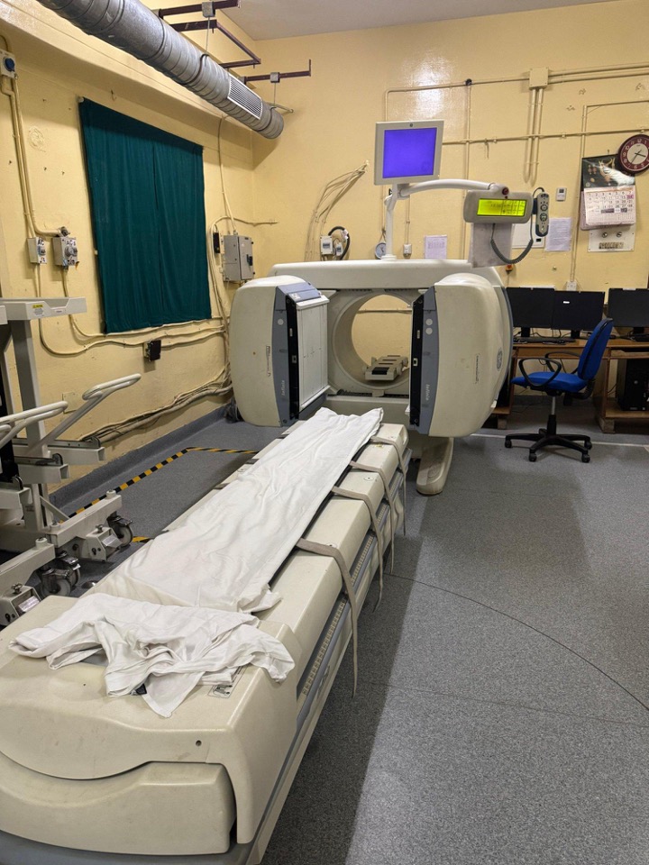
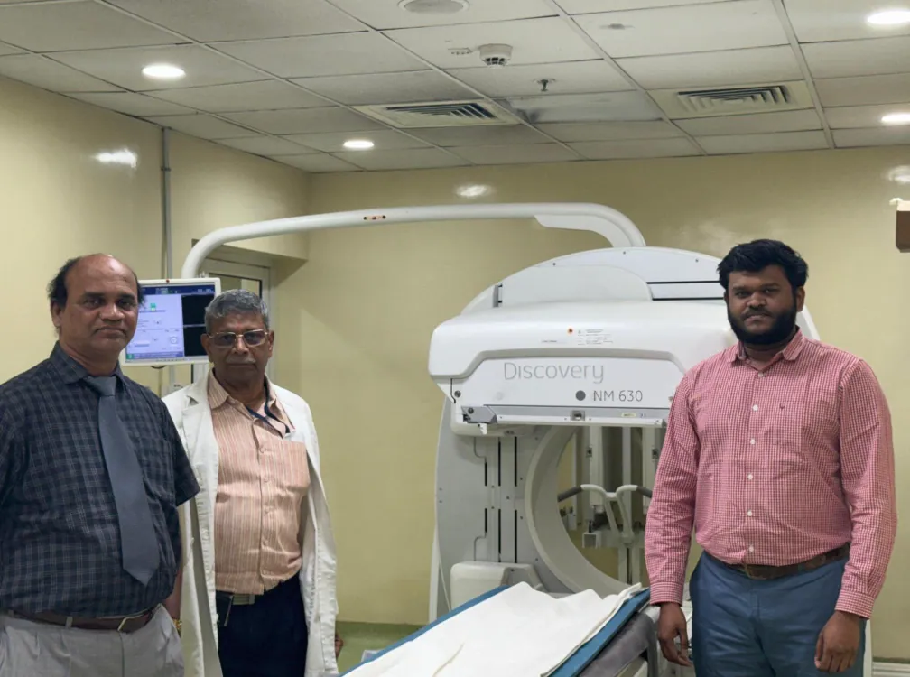
Advanced dual-head gamma camera with SPECT capability for high-quality nuclear imaging of all organs
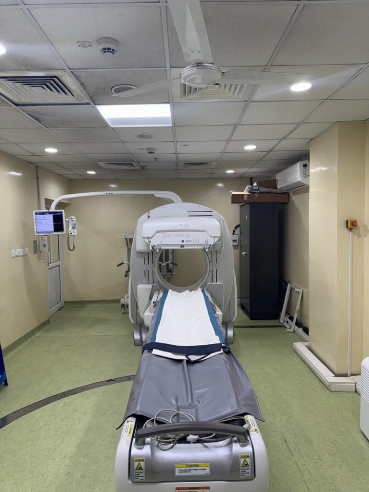
State-of-the-art nuclear medicine imaging system for precise diagnostic scans
Analyzes energy spectrum of gamma radiation for quality control and radiopharmaceutical purity testing
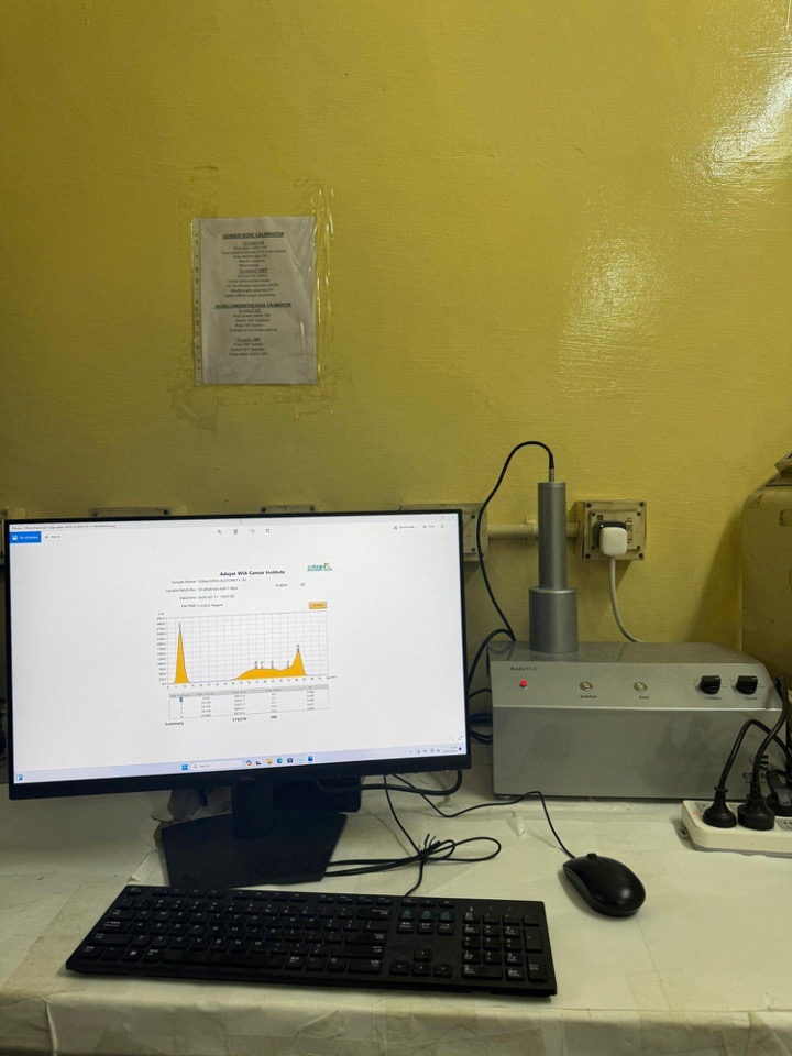
Instant Thin Layer Chromatography system for radiopharmaceutical quality assurance
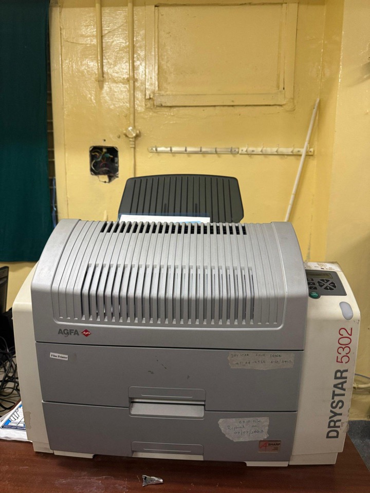
High-quality medical image printing systems for producing diagnostic-grade hard copies of nuclear medicine scans
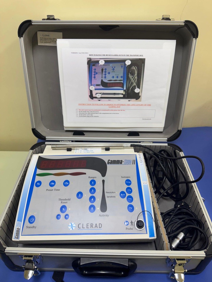
Intraoperative radiation detection device used during sentinel lymph node biopsy and tumor localization surgery
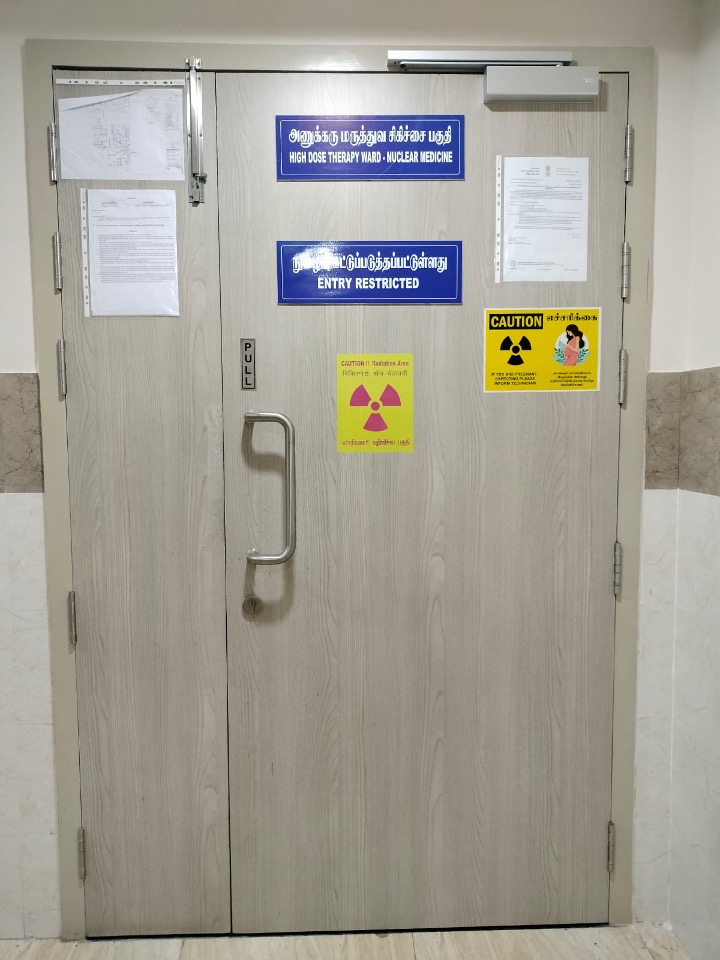
Dedicated isolation facility with proper radiation shielding for radioiodine therapy and other therapeutic nuclear medicine procedures
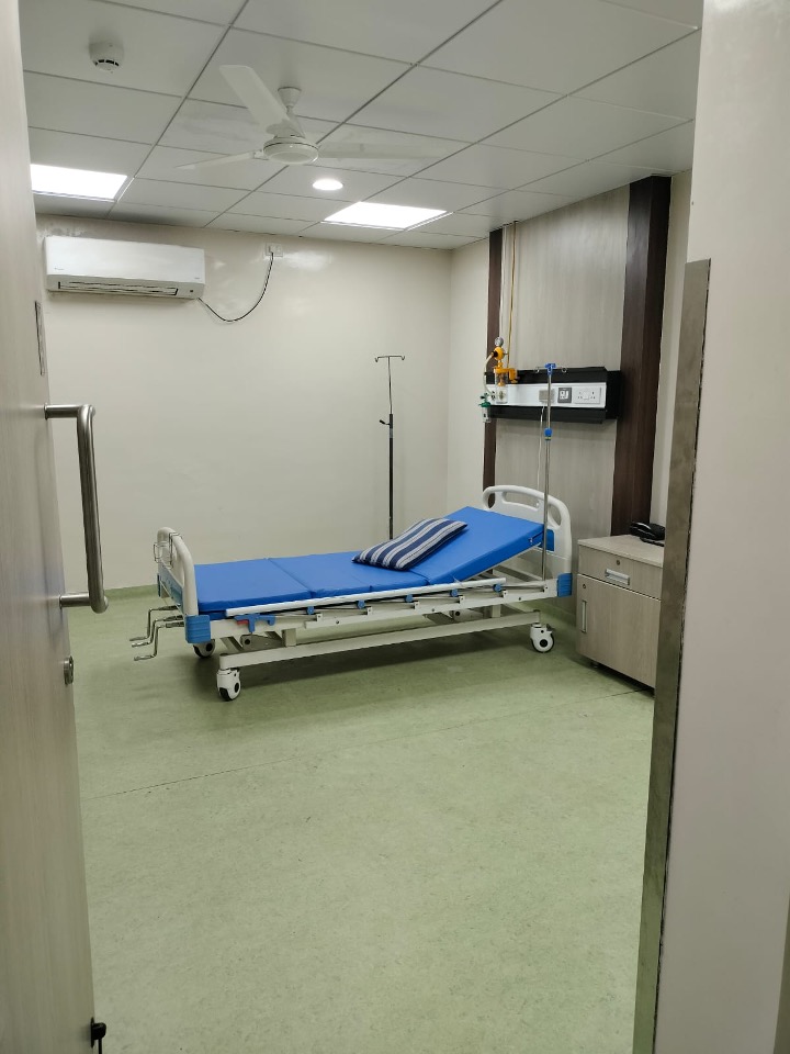
Comfortable patient rooms equipped with all necessary amenities and radiation safety features
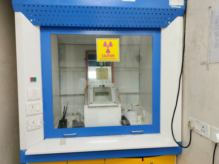
Ventilated enclosure for safe handling of radioactive materials in patient care areas
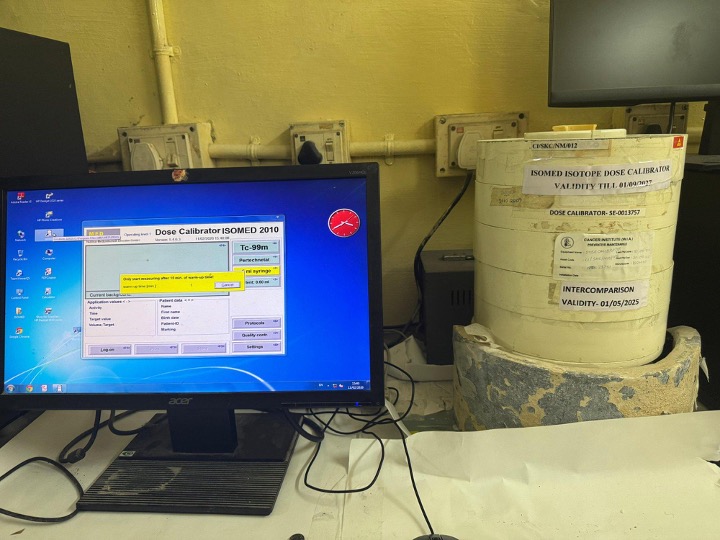
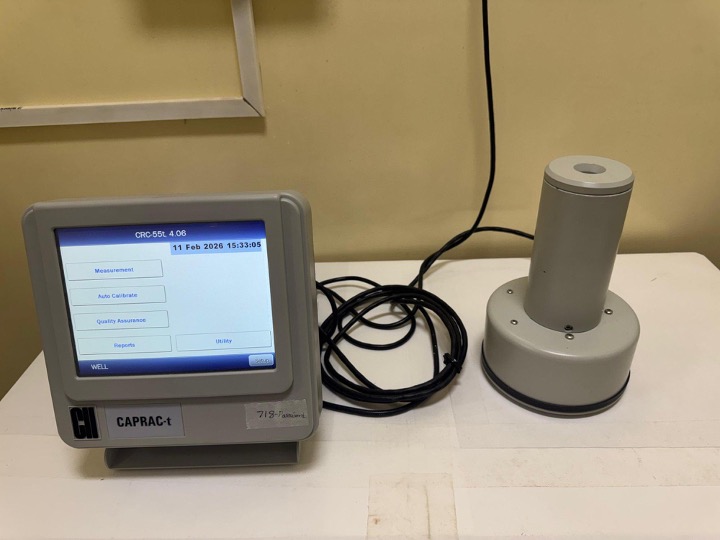
Precisely measures radioactivity of radiopharmaceuticals before administration to ensure accurate patient dosing
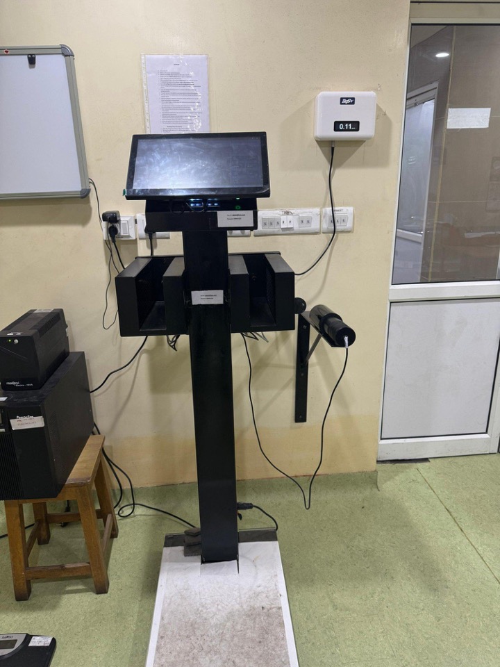
Detects radioactive contamination on hands and feet of staff and patients for safety compliance
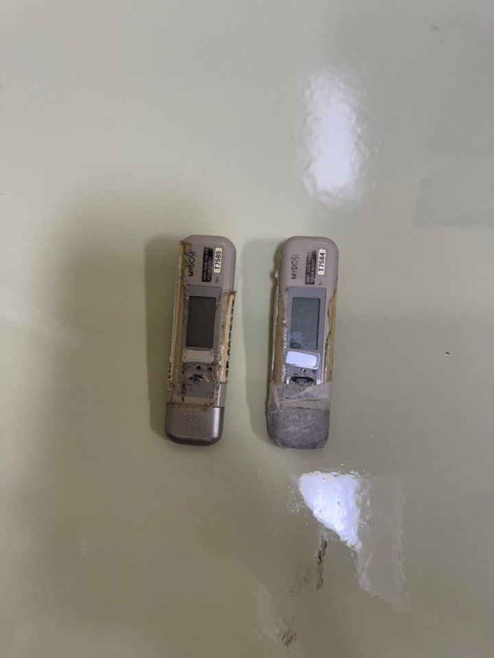
Personal radiation monitoring device worn by staff to track individual radiation exposure levels
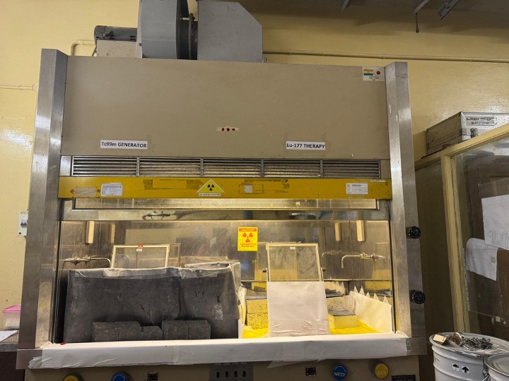
Laminar flow workstations with LED lighting for safe handling and preparation of radiopharmaceuticals
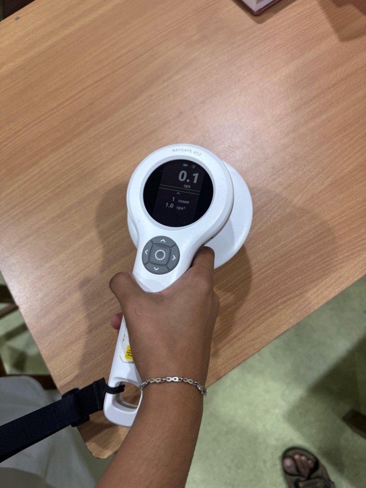
Detects and measures radioactive contamination on surfaces and equipment
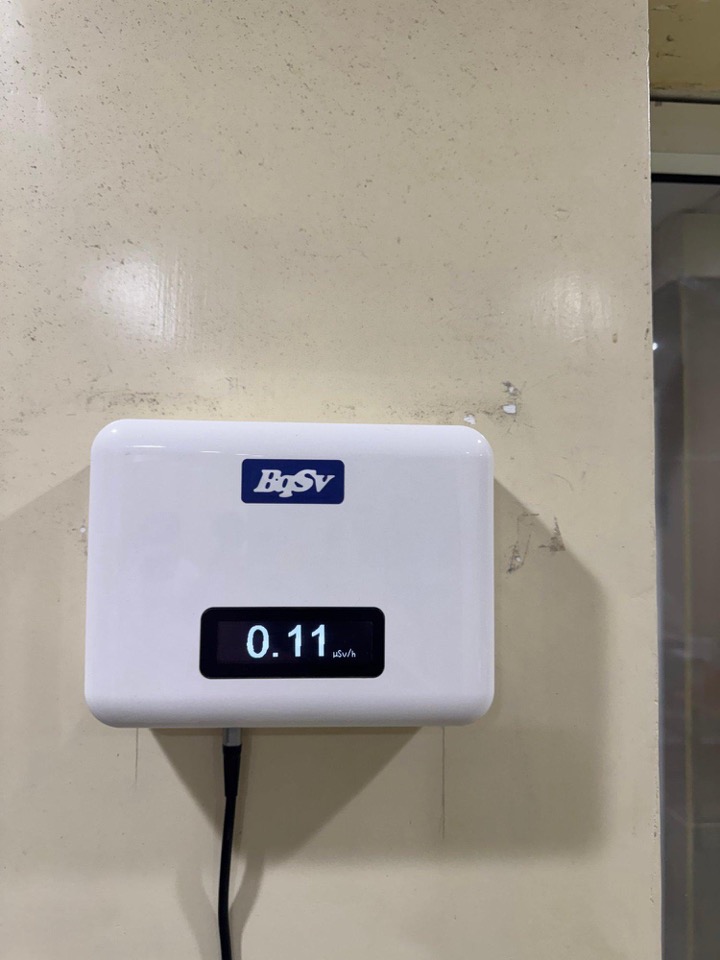
Continuously monitors ambient radiation levels in the department to ensure staff and patient safety
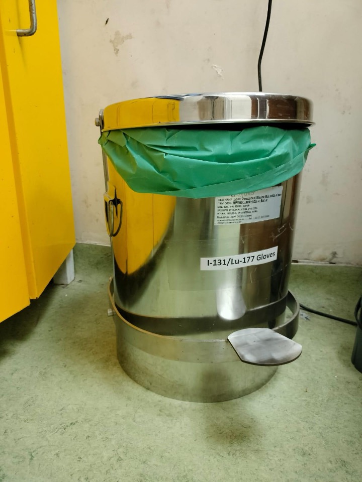
Shielded containers for safe disposal of radioactive waste materials
Personal protective equipment providing radiation shielding for staff during procedures
Technologist processing nuclear medicine images for diagnostic interpretation
Patient guidance
Yes. The doses used are carefully chosen to provide the maximum diagnostic information with the minimum radiation exposure. Most PET/CT scans deliver a radiation dose similar to a few months of natural background radiation. The radiotracer decays and is eliminated from the body within hours to days.
It depends on the scan. FDG PET/CT requires 4-6 hours of fasting to ensure low blood glucose, which maximises tracer uptake in tumours. Bone scans and most SPECT studies do not require fasting. Your specific preparation instructions will be confirmed when you are booked.
For most scans, yes. Exceptions include brain scans where sedation may be used. We recommend drinking plenty of water after your scan to help clear the tracer through your kidneys.
Please inform us if you are pregnant or breastfeeding, have diabetes, are claustrophobic, or have had a recent scan or surgery. Also bring a list of your current medications, as some may need to be held before certain studies.
Allow 2-3 hours for most PET/CT scans, including the uptake period. SPECT/CT scan requires 3-4 hours. Your appointment letter will confirm the expected duration.